")
Surgical techniques for a good outcome in cataract surgery: personal perspectives
Related content
The mandate of this article is to provide practical examples of how to achieve safe, good quality cataract surgery with different surgical techniques.
Cataract outcomes and case selection
In India, increasing pressure to clear cataract backlogs has placed emphasis on number only. If outcomes and quality are ignored, we not only convert curable blindness to incurable but also create adverse publicity for our programme. Case selection is an important issue. In this context, it is important to appreciate that a torch light examination alone may not be sufficient to detect pre-existing pathology (e.g., glaucoma or macular degeneration) that can contribute to poor visual results. Signs, such as mild subluxation of the lens, detected on a slit lamp can result in a change of surgical plan from ECCE to ICCE.
Complications
Wound related prob lems, endothelial damage, vitreous loss and post-operative infections are the common factors that contri bute to poor outcomes in any type of cataract surgery. Good surgical technique and appropriate management of complications can help minimise these.
Anaesthesia
A soft, well anaesthetised eye is vital to the success of intra capsular (ICCE) and standard extra capsular cataract surgery (ECCE). Use of hyal uro nidase and intermittent digital pressure (released every 30 seconds) spreads the anaesthetic and reduces the vitreous volume for safe surgery. Though the modern small incision cataract surgery (both phacoemulsification and manual) can be performed under topical anaesthesia, peribulbar injections may be more suited for the average surgeon.
Lighting and magnification
Good lighting and magnification improves visibility and is required even for ICCE. A good microscope fulfils these two requirements. Wound construction, recognition and management of problems like residual cortex, posterior capsule rupture, vitreous loss, etc, are all better dealt with using a good quality microscope.
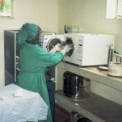
Sterilization and post-operative infection
Commensal bacteria in and around the eye may be the cause of endophthalmitis. Cleaning the periorbital skin including the eyebrows and eyelids with povidone iodine will reduce the bacterial load. In order to increase contact time, this is done at the time of anaesthesia as well as prior to surgery.
Povidone iodine skin preparation (Beta dine) should be used as a 5% solution. Povidone iodine eye drops can have strengths of 2.5% or 1%. The skin preparation and eye drops should not be confused. Instruments should only be sterilized in appropriate chemicals (see Ingrid Cox and Sue Stevens with references: pp. 40-41) and always thoroughly rinsed and flushed through with sterile water before use.
No-touch technique
The ‘no-touch’ technique is effective in minimising contamination. In the no-touch technique both the surgeon and the assistant ensure that only tips of the instruments make contact with the eye and do not touch anything else.
Surgical blades and wound size
Use of a sharp blade ensures a clean wound that will appose well. Being ‘poor mission hospital doctors in a poor developing country’ we re-sterilize and re-use expensive blades. However, beyond a certain number of ‘uses’, this is actually detri-mental to the outcome. The money saved may not be worth it.
As far as the wound size is concerned, for ICCE and ECCE it is better to err on the side of a larger wound. For ICCE this helps in adequate retraction of the cornea for cryoextraction and helps prevent capsular rupture. In the usual manual ECCE the larger wounds facilitates the nucleus expression; and excess pressure that can result in zonular dialysis or posterior capsular rupture is avoided.
Use of an anterior chamber maintainer (ACM)
Maintaining a formed anterior chamber (AC) during surgery can minimise endothelial loss. This is usually achieved by the use of methyl cellulose, or air. We use an ACM. In our hands, the ACM (Fig.1) is an excellent tool for use in small incision surgery, both manual and with phacoemulsification. It is usually introduced at the very beginning of surgery and has several advantages. The ACM keeps the anterior chamber deep during capsulotomy. It firms up the eye and facilitates the incision. All the flow is outwards: debris is washed out and contamination is decreased. Fluid lost from the eye is immediately replaced; turbulence is reduced and the IOP is maintained. Theoretically, the constant IOP should decrease the risk of expulsive haemorrhage. A well formed AC protects the endothelium and assists rotation of the nucleus where this is required. The fluid under pressure helps complete hydro dis sec tion. In the manual small incision technique that we use, the ACM flow is also used to express the nucleus.
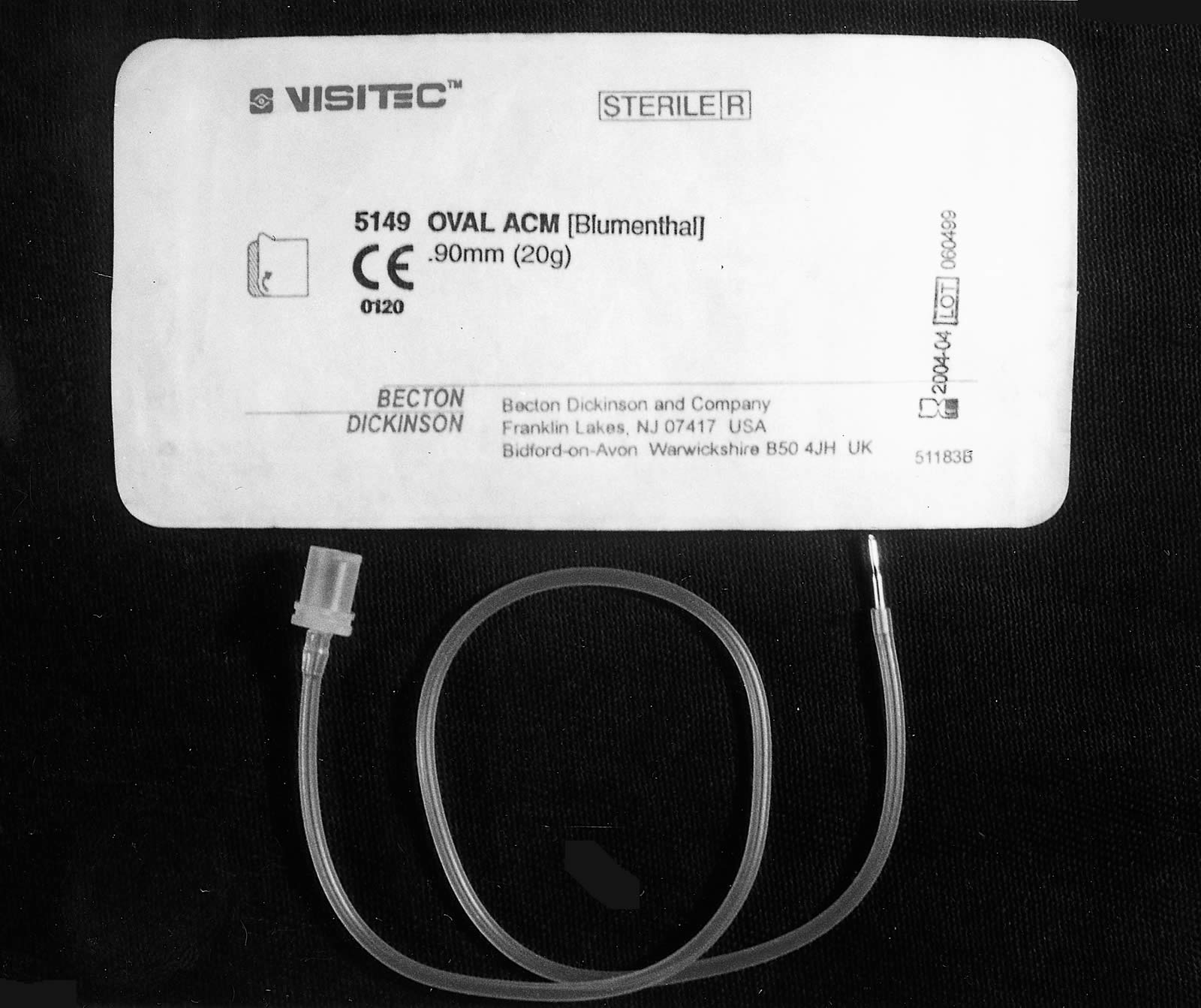

Cortex is safely aspirated through a paracentesis with a syringe attached to a cannula; this is done in a deep and stable anterior chamber provided by the ACM (Fig. 2). We use this technique for cortex aspiration even with phacoemulsification. In the event of vitreous loss, an ACM facilitates the vitrectomy. We believe in the ACM and strongly recommend its use routinely, as well as for teaching small incision surgery.
Vitreous loss and vitrectomy
Vitreous loss is the most common complication of cataract surgery; if not managed appropriately it can cause delayed and irreversible visual loss. We recommend a mecha nised vitrector to manage this problem. The objectives of a vitrectomy for vitreous loss are:
- Avoid vitreous incarceration into the wound.
- Avoid vitreous contact with other anterior chamber structures like the corneal endothelium and iris.
The height of the ACM is lowered and the vitrector is introduced through the para-centesis. Cutting and aspiration is initiated within the region of the posterior capsular tear, thereby removing vitreous from the chamber and wound. A second paracen-tesis is used to introduce a cannula that sweeps the vitreous away from the wound. If an ACM is not used, this paracentesis serves to provide irrigation through a cannula. If available, a light pipe (endo-illuminator) is useful to determine the presence of residual vitreous and confirm adequate capsular support for the IOL.
Wound closure
As far as wound closure is concerned, it is the correct apposition and not the number of sutures that is important. Pre-placed marks across the line of the wound and using irregularities in the wound like a jigsaw puzzle help in proper apposition. Using the circular ring at the end of a safety pin as a simple per operative kerato meter and keeping a large air bubble in the AC while suturing prevents high astigmatism.
We hope this article has provided some practical guidelines that will help improve outcomes. We realise that we may have raised more questions than have been answered, but space constraints prevent detailed discussion of techniques.