")
Training in trichiasis surgery
Related content
Introduction
It is estimated that each village in central Tanzania has between 5 – 25 persons with in-turned eyelashes due to trachoma. Half of these people constantly epilate their eyelashes to ease the irritation and pain from in-turned eyelashes.

Aims
The aims of training in trichiasis surgery are to teach:
- Identification of patients needing trichiasis surgery
- A good and safe surgical procedure
- The principles and practice of competent follow-up.
Selection of trainees
Trainees are recommended by their respective Health authorities. They are required to have:
- Previous experience in eye examination
- Experience in giving injections
- Knowledge of sterile surgical techniques
- Previously observed eye surgery.
Two weeks is the minimum time recommended to train a trichiasis surgeon.
Objectives of training in trichiasis surgery
At the end of the course the trainee should be able to:
- Perform the tarsal rotation method for trichiasis
- Complete at least 5 supervised operations to receive certification
- Follow-up trichiasis patients and recognise any complications
- Complete reports and keep records of trichiasis surgery
- Assess competence and improve surgical skills, under supervision
- Recognise the barriers to trichiasis surgery and how these can be overcome
- Assist in the planning and implementation of mobile eye clinics (community-based)
- Demonstrate trachoma assessment methods
- Demonstrate skills in trachoma grading
- Implement SAFE interventions as part of comprehensive eye care.

Handouts
Handouts on the following topics, to support teaching sessions, are distributed to participants during the training period:
- Primary Health Care.
- The 8 Elements of Health Care.
- The 5 Principles of Primary Health Care.
- Anatomy of the Eye (main emphasis on the upper eyelid).
- Checklist of Supplies and Materials.
- Sterilization Methods.
- Record Keeping.
- The SAFE Strategy.
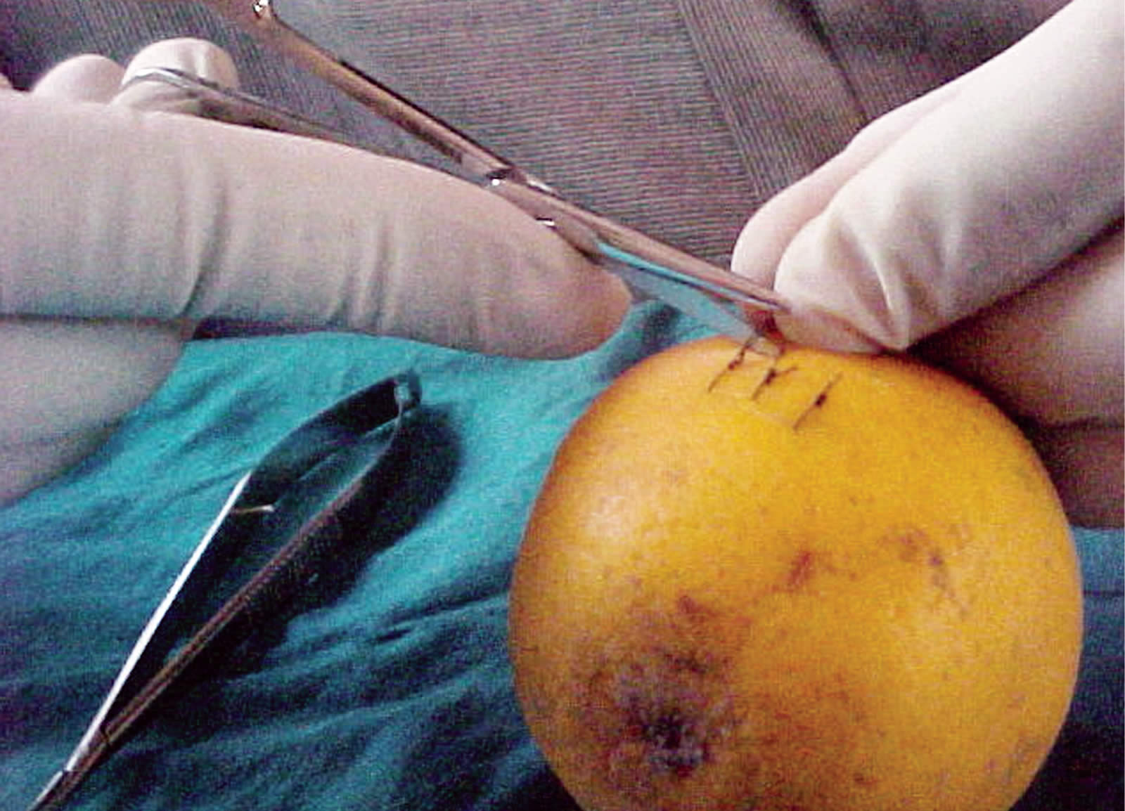
Incision and stitching exercises
The trainer demonstrates the procedure. It is then practised on oranges and bananas.
The steps include:
- Incising the ‘eyelid’-orange peel or banana
- Everting and incising the ‘conjunctiva and tarsal plate’-inside of orange peel
- Completing the incision with scissors
- Suturing the ‘eyelid’.
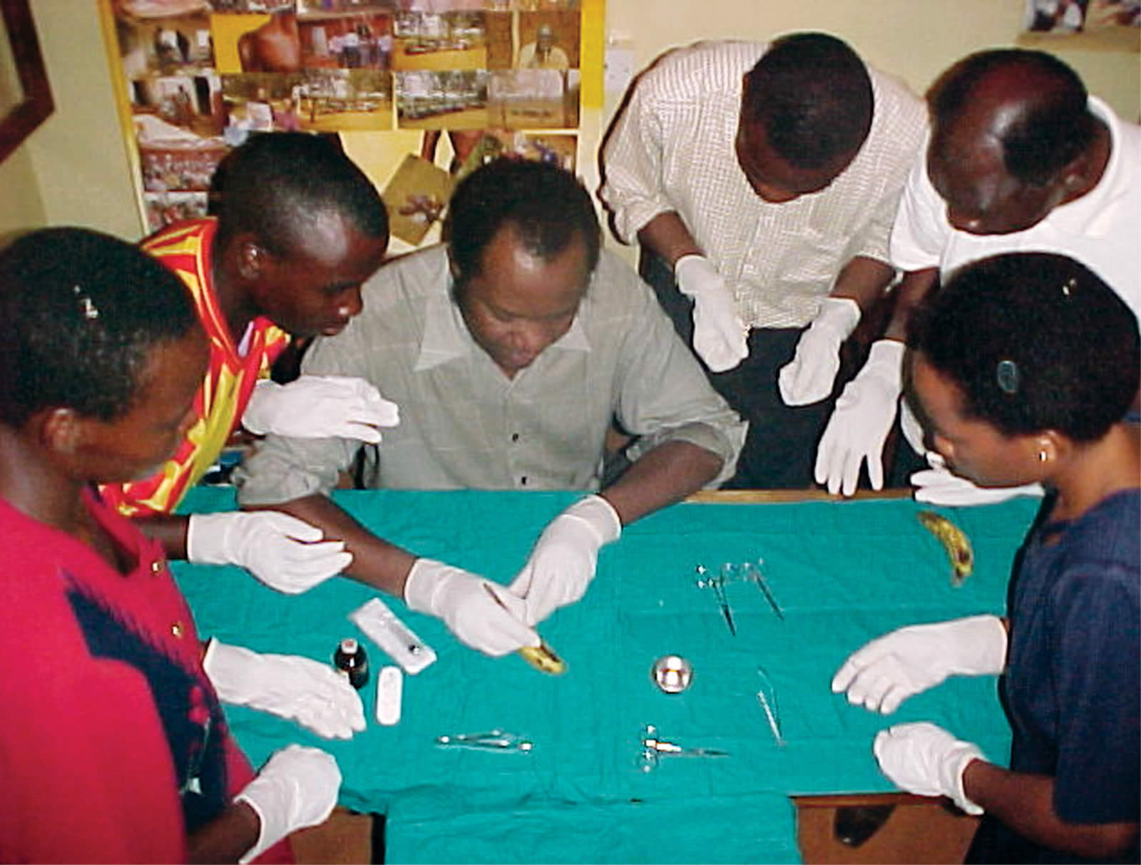
Handling surgical instruments
The key skills in which each participant must be competent are:
- Holding the needle holder in the dominant hand
- Mounting the needle (with suture) on the needle holder
- Making sure the needle holder holds the needle one-third away from the tip
- Holding the toothed dissecting forceps with the other hand
- Holding the edge of the distal skin edge and inserting a needle with suture
- Holding the skin on the proximal side, and pushing the needle through the skin
- Pulling the suture and making the first, second, and third knots
- rutting the two ends of the suture material (about 0.5mm long above the wound; this helps when removing sutures).
After everyone has successfully mastered incision and suturing skills, they each receive their own surgical kits for the field visits.
The trainer reviews the schedule for the field visits and reviews the sterilization procedures for surgical kits during the visits and at the end of each day.
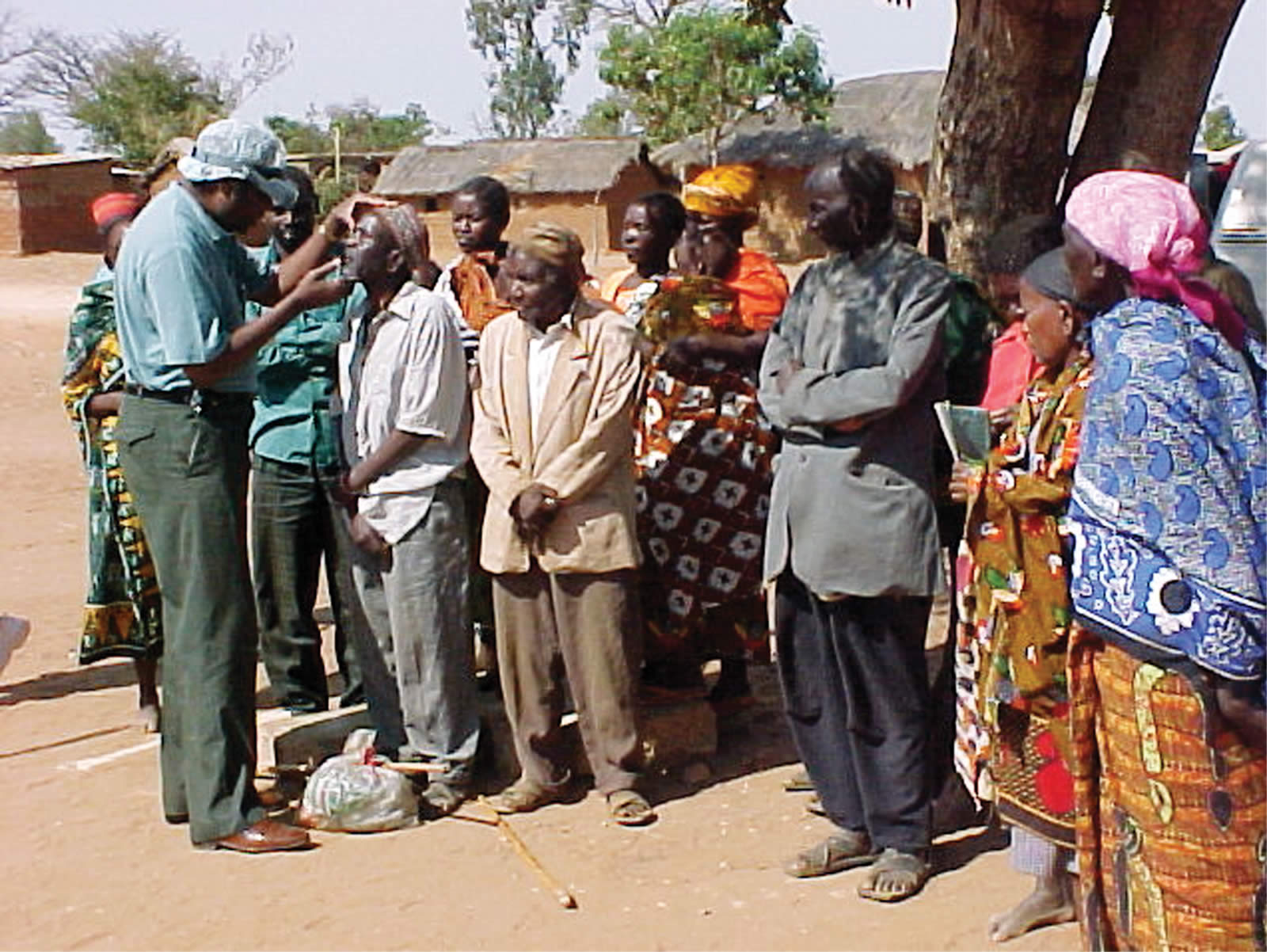
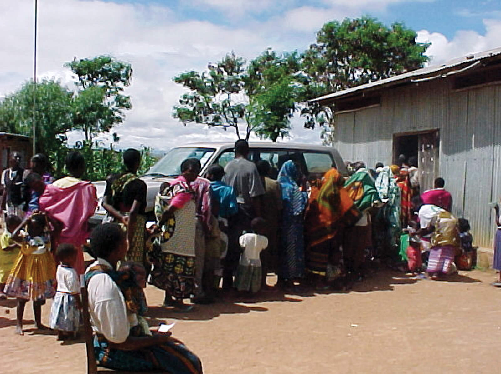
Mobile eye clinic: procedure for trichiasis patients
During the mobile eye clinic, priority should be given to:
- patients with trichiasis
- people who are blind
- patients with painful red eyes.
Patients with trichiasis are sent immediately for visual acuity testing. These patients are then guided to the operating area. The outpatient form is carried with the patient into the operating area. Numbers are written on the outpatient form so that surgical teams will know how many patients to expect.
The following procedures are followed:
- Identify who will take the patient home and make sure all the procedures are understood, the need for return in 7 days clearly stated, and verbal consent given
- Written consent is advised
- Before the patient lies on the operating table, the surgical team checks the fitness of the patient for surgery (checking blood pressure, allergies to drugs, shortness of breath, heart problems, mental state )
- The patient is asked if they have consumed any alcohol that day. If so, trichiasis surgery should be postponed
- Community health workers and/or the advance team should advise patients of this requirement during the preparation screening, before the days of the clinic
- After surgery, the patient takes the outpatient form and follow-up requirements are explained to the patient.
Normally, trichiasis surgery is performed on one eye at a time. The second operation can take place when the sutures of the first eye are removed after eight days. Some patients may choose to delay the second operation until the first eye is completely healed.
The trichiasis surgical procedure: Bilamellar Tarsal Rotation Procedure (BTRP)
With sterile instruments and other supplies at hand, the procedure should be done following these steps:
Preparations:
a) Clean the skin surrounding the eye with an antiseptic solution.
b) Amethocaine eye drops instilled on the eye (or similar topical anaesthetic).
c) Scrub the hands with soap and water for at least 5 minutes.
d) Put on sterile gloves (gloves must be worn).
e) Clean the patient’s face and eyes.
Surgical procedure:
a) A local anaesthetic injection is given into the upper lid (ask the patient to look down).
b) Usually 3 mls are sufficient (never inject more than 5 mls in any single operation).
c) An operation is performed seated at the head of the patient.
d) For better visibility, a magnifying loupe is used and a flashlight held by an assistant.
e) The eyelid is ‘fixed’.
f) The upper eyelid is incised (incision of the skin and muscle must be parallel to the lid margin and 3 mm above it, the entire distance between the haemostats).
g) Eversion of the eyelid is then done.
h) Incision of the conjunctiva and tarsal plate, through its full thickness, parallel to the lid margin and 3mm above it, the entire distance between the haemostats.
i) The incision is then united by inserting the points of the closed scissors into the incision in the conjunctival-tarsal plate, through remaining intact muscle, and out through the skin-muscle incision.
j) Open the scissors while still held across the lid: the blunt aspect of the blades will spread apart intact muscle-repeat until it is a full thickness hole.
k) Remove the haemostats.
l) Complete the incision medially and laterally using the scissors.
m) Suturing the eyelid is then done using 4/0 silk or chromic catgut. This is to re-attach the distal fragment in an outwardly rotated position, so that the eyelashes no longer rub on the cornea. This is achieved by anchoring sutures on the conjunctival surface of the proximal fragment, and running them over the distal tarsal plate to exit through the skin near the eyelashes, thus drawing the lash margin outwards and upwards.
Summary of follow-up care
Day 1: Lid surgery patients advised of time to return for follow-up care.
Day 2: Patient returns to meet health worker, who removes eye patch, cleans wound and applies tetracycline eye ointment. Any patient with excessive bleeding, swelling and/or severe pain should be referred immediately.
Day 3-7: Patients continue to see health worker on daily basis – to have the wound cleaned and to have tetracycline eye ointment applied.
Day 8: Mobile eye team returns – sutures are removed.
At the end of the trichiasis operation day, the principal trainer reviews follow-up procedures for patients who have undergone community-based trichiasis surgery with community health workers or their equivalents. Each community health worker should receive a list of the trichiasis patients. Each patient is given tablets of paracetamol to be taken as needed for pain relief, once in the morning and once in the evening.
Overcoming fears
Patients who are afraid of having trichiasis surgery receive counselling. Fear of the injection, cutting, pain and bleeding are most often the concerns expressed. Patients are asked to speak to someone who has had the operation and will talk about his/her experience.
Constraints to surgery
- People do not know that this is a problem that can be solved.
- People are afraid of the operation.
- Communities lack transport to take patients to the hospital or clinic.
- The number of people who are able to do the surgery is limited.
- Bad service by service providers, e.g., bad language, etc.
- The cost of surgery in some hospitals is not affordable to most patients.
Graduation and certification
Each participant is given:
- A certificate after successfully performing the operations – under supervision.
- A copy of the WHO Manual ‘Trichiasis Surgery for Trachoma – The Bilamellar Tarsal Rotation Procedure’.
- 100 tubes of tetracycline.
- 4 sutures, a surgical set for minor operations, and other supplies as available.
- A set of bi-monthly reporting forms to be used within their health system.
Example of a trichiasis register
| Date | No. | Name | Sex | Address | VR | VL | TT situation | Remarks |
|---|---|---|---|---|---|---|---|---|
Further reading
1 Johnson G J, Minassian D C, Weale R. The Epidemiology of Eye Disease 1998. pp 132-135.
2 Reacher M, Foster A, Huber J. Trichiasis Surgery for Trachoma: The Bilamellar Tarsal Rotation Procedure. WHO/PBL 1998.
3 Ballen P H. A simple procedure for the relief of trichiasis and entropion of the upper lid. Arch Ophthalmol 1964; 72: 239 – 240.
4 Reacher M H, Munoz B, Alghassany A, Daar A, Elbualy M, Taylor H. A controlled trial of surgery for trachomatous trichiasis of the upper lid. Arch Ophthalmol 1992; 110: 667- 674.